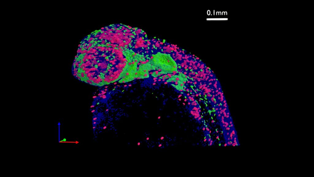
zebrafish embryo - 36 hours post fertilization
sketchfab
Scientists skillfully tag proteins produced by cells with fluorescent markers, enabling them to track these proteins as well as the cells that carry them, and study their activity in detail. On this image, researchers successfully track neural crest cells by tagging a protein exclusively produced by this cell type, marked in green, indicating sox10. They also examine proliferating cells by tagging a protein only made by cells undergoing division, highlighted in red, representing phospho-histone H3. Meanwhile, the DNA of every cell is stained with a blue chemical, clearly revealing the nuclei. By combining these different elements of observation, scientists can rigorously test their hypothesis and draw accurate conclusions.
With this file you will be able to print zebrafish embryo - 36 hours post fertilization with your 3D printer. Click on the button and save the file on your computer to work, edit or customize your design. You can also find more 3D designs for printers on zebrafish embryo - 36 hours post fertilization.
