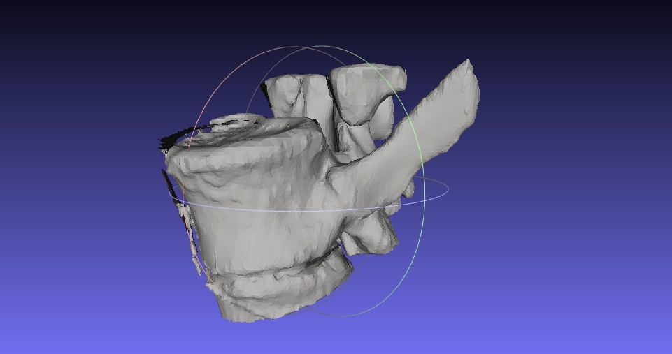
Vertebrae (MRI)
thingiverse
The human spine is comprised of 33 individual bones known as vertebrae, which are stacked on top of one another to form the spinal column. These bones are separated by intervertebral discs, which act as shock absorbers and help to distribute weight evenly throughout the body. In a normal MRI study, the vertebral bodies are visible as rounded or oval-shaped structures that appear bright white due to their high water content. The intervertebral discs are seen as dark areas between the vertebrae, while the spinal cord is visible as a thin, grayish-white structure running along the length of the spine. The MRI scan can also reveal any abnormalities in the vertebral bodies or intervertebral discs, such as fractures, herniated discs, or degenerative disc disease. For example, a fractured vertebra may appear as a darkened area within the affected bone, while a herniated disc may show up as a bulge or protrusion into the spinal canal. In addition to visualizing the vertebral bodies and intervertebral discs, MRI studies can also help diagnose conditions such as spinal stenosis, spondylolisthesis, and scoliosis. These conditions occur when the vertebrae become misaligned or develop abnormal growths, which can put pressure on surrounding nerves and cause pain, numbness, or tingling sensations in the arms and legs. Overall, MRI studies provide valuable information about the health of the spine and can help doctors diagnose a wide range of spinal disorders. By analyzing the images obtained from an MRI scan, medical professionals can develop effective treatment plans to alleviate symptoms and prevent further damage to the vertebral bodies and intervertebral discs.
With this file you will be able to print Vertebrae (MRI) with your 3D printer. Click on the button and save the file on your computer to work, edit or customize your design. You can also find more 3D designs for printers on Vertebrae (MRI).
