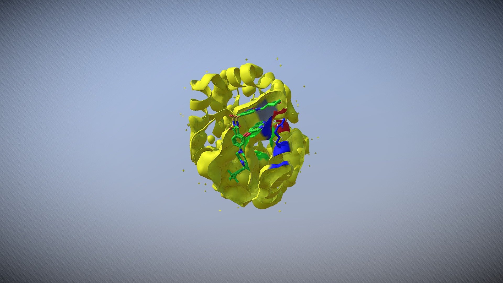
The structure of BCL-2 with Venetoclax
sketchfab
BCL-2 proteins act as master regulators controlling cell death processes. The BCL-2 protein structure boasts two hydrophobic α-helices flanked by 6-7 amphipathic α-helices, creating a unique molecular framework. A critical binding site for proapoptotic proteins emerges from four of the amphipathic helices, forming a hydrophobic groove that facilitates protein interactions. Moreover, BCL-2 proteins possess a potent BH3 domain, crucial for mediating protein-protein interactions and influencing cell fate decisions. The interactions in P2 and P4, two hydrophobic pockets, and electrostatic interactions between aspartic acid and arginine residues on proapoptotic and antiapoptotic proteins govern the binding to BCL-2 proteins. The P2 pocket is a critical determinant of BH3 peptide selectivity. Adjacent residues A100 and D103 define the boundaries of the P4 pocket, while the F104 side chain separates the P2 and P4 pockets of BCL-2. Additionally, the nitrogen atom of the azaindole forms a hydrogen bond with Asp103 of BCL-2, reinforcing protein interactions.
With this file you will be able to print The structure of BCL-2 with Venetoclax with your 3D printer. Click on the button and save the file on your computer to work, edit or customize your design. You can also find more 3D designs for printers on The structure of BCL-2 with Venetoclax.
