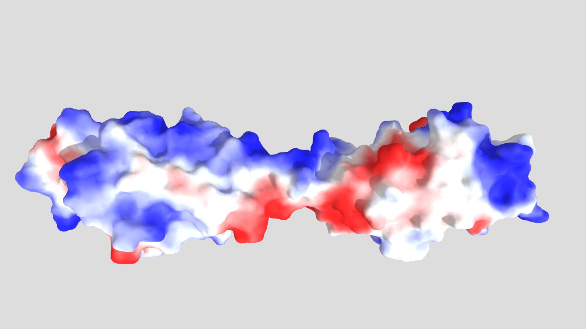
Myotilin_F-actin_Fig2B-Ig1_Ig2_electrostatic pot
sketchfab
Surface electrostatic potential mapped onto the myotilin Ig1Ig2 structural model reveals distinct patterns of charge distribution, as illustrated in the immunoglobulin-like domains. Colored surfaces showcase a gradual shift from deep red to pure white and finally blue, reflecting calculations performed by the Adaptive Poisson-Boltzmann Solver software.
Download Model from sketchfab
With this file you will be able to print Myotilin_F-actin_Fig2B-Ig1_Ig2_electrostatic pot with your 3D printer. Click on the button and save the file on your computer to work, edit or customize your design. You can also find more 3D designs for printers on Myotilin_F-actin_Fig2B-Ig1_Ig2_electrostatic pot.
