Myotilin F-actin Fig1E-EOM Models Trx- Myot
sketchfab
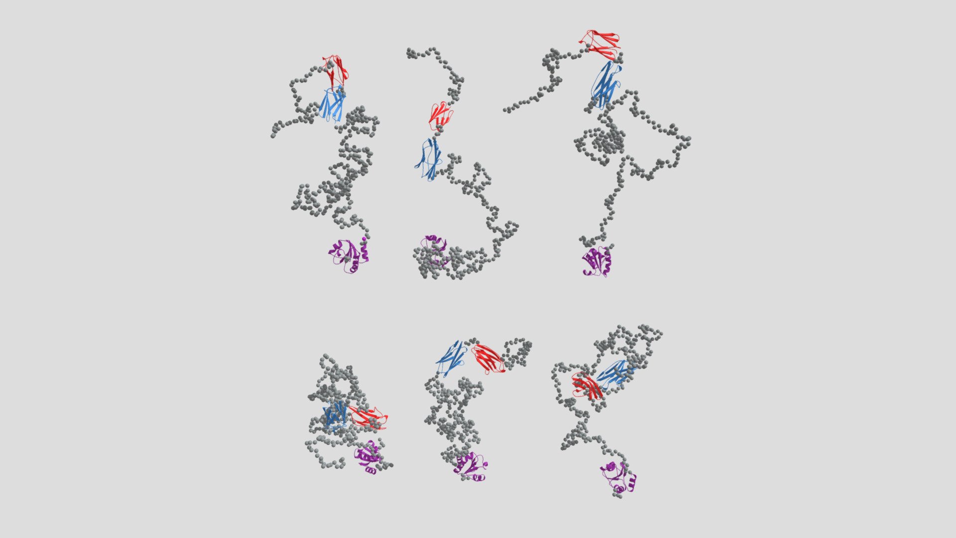
SAXS derived ensemble optimization methods models of sarcomerox Z-disc protein myotolin in chimera with thioredoxin (Trx-MYOT) reveal flexible and intrinsically disordered N- and C-terminal regions, depicted in grey. The chosen models are displayed with decreasing percentage contribution, calculated from the final population of SAXS derived ensemble optimization methods models. The N-terminal section of myotilin contains a serine-rich region, which comprises a hydrophobic residues stretch (yellow) and represents a "mutational hotspot" of the protein, highlighted in grey. Known disease-causing mutations are indicated. C-terminal immunoglobulin domains 1 and 2, colored blue and red respectively, are followed by the C-terminal tail.
With this file you will be able to print Myotilin F-actin Fig1E-EOM Models Trx- Myot with your 3D printer. Click on the button and save the file on your computer to work, edit or customize your design. You can also find more 3D designs for printers on Myotilin F-actin Fig1E-EOM Models Trx- Myot.
