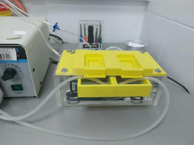
Interface chamber for brain slice electrophysiology
prusaprinters
This is an interface chamber for brain slice electrophysiology. With acute rodent hippocampal slices incubated for 3 hours in this interface chamber sharp wave ripples can be reliably recorded. The “aquarium” part of the interface chamber has an aquarium heater that can heat up to 34 degrees celcius. The top part is attached via screwes ( it doesn't have to be airtight, don't worry). The collector parts collect ACSF that is recycled using a peristaltic pump. The “wall” part is used to close the open front part of the chamber, and on top you should cover each side with a recgangular piece of plastic covered in tissue so that condensation doesn't drip onto the slices. Carbogen is delivered via a silicon tube with lots of small perforations. The ACSF for perfusion is delivered via a 21g needle that has had the end cut off. It is attached to a tube that is submerged in the “aquarium” so that the ACSF is heated. The slices lay on top of two layers of lense cleaning tissue, and each slice is on its own extra square of lense tissue. Strips of material (small parts cur from women's tights are ideal) are used to provide flow past the slices.After incubation slices should be transferred (by picking up the smal square of lense tissue with foreceps) into a double perfusion submerged chamber and perfused with ACSF at a rate of 9–10 ml/min at 32 ± 1°C.
With this file you will be able to print Interface chamber for brain slice electrophysiology with your 3D printer. Click on the button and save the file on your computer to work, edit or customize your design. You can also find more 3D designs for printers on Interface chamber for brain slice electrophysiology.
