Influenza Virus Infecting Cells
sketchfab
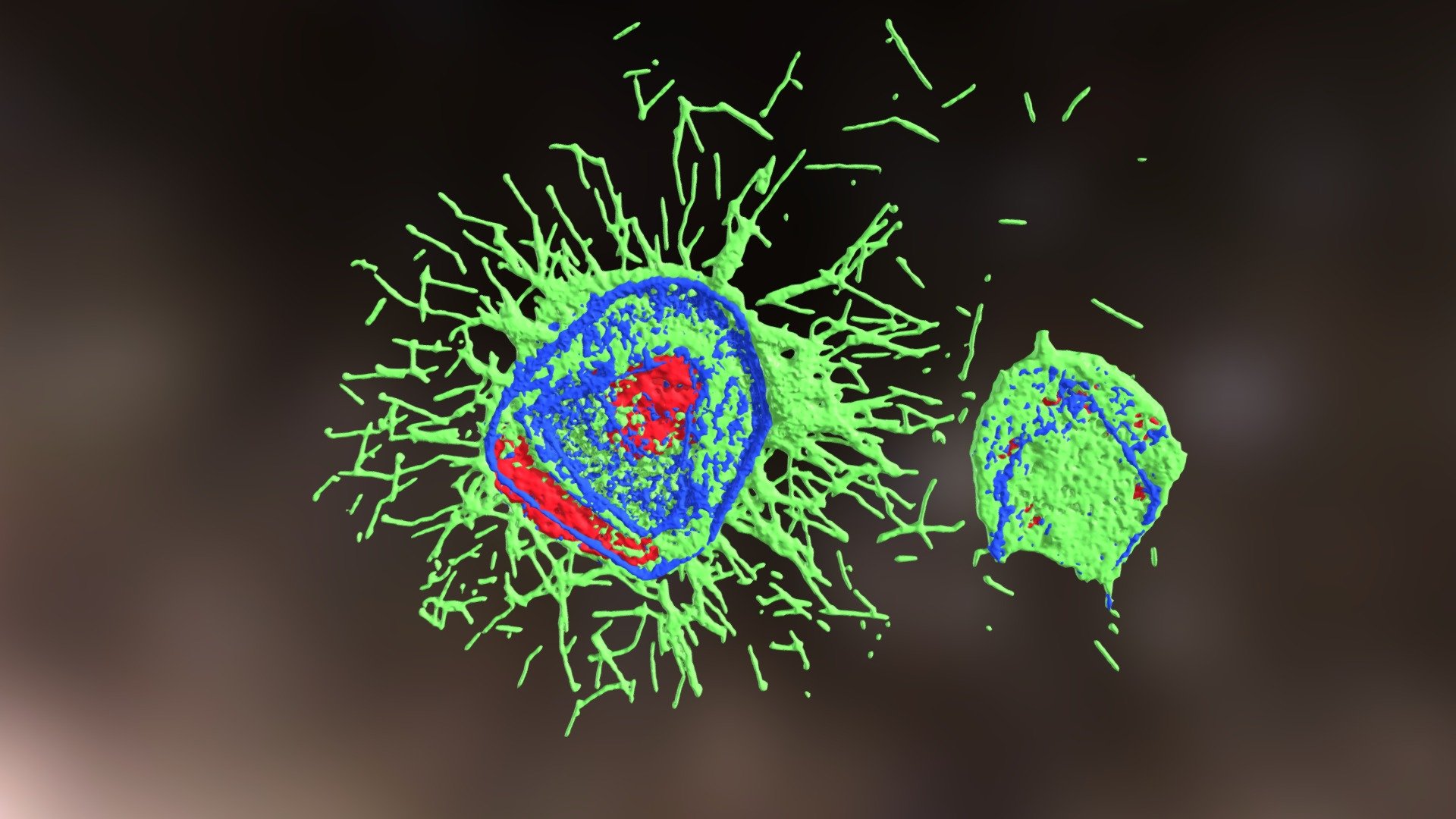
Human cells infected with Influenza Virus H3N2 Udorn strain display distinct characteristics under microscopic examination. Hemagglutinin protein, labelled with Alexa 488 dye, is clearly visible in green, highlighting the formation of viral filaments as they bud from the host cells. Meanwhile, actin, marked by Phalloidin-Alexa 568, appears in vibrant red hues, while the DAPI-stained nuclei shine in brilliant blue tones. A Zeiss LSM-710 confocal microscope captured the intricate details of this process with precision, providing valuable insights into the viral replication cycle. Microscopic data was generously provided by Colin Loney at the MRC Centre for Virus Research.
With this file you will be able to print Influenza Virus Infecting Cells with your 3D printer. Click on the button and save the file on your computer to work, edit or customize your design. You can also find more 3D designs for printers on Influenza Virus Infecting Cells.