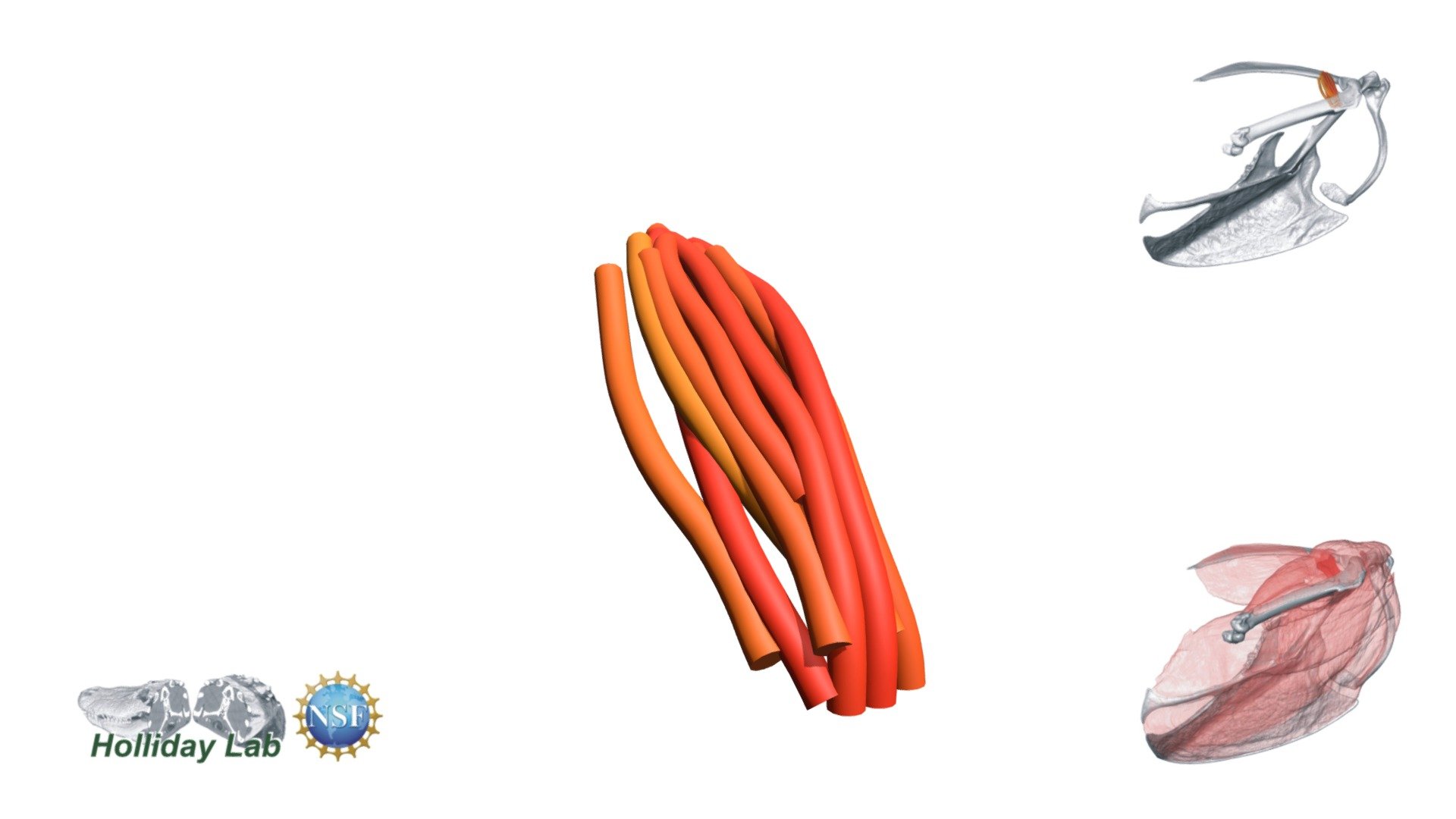
Figure 20. Starling scapulohumeralis cranialis
sketchfab
The detailed anatomical structure of the m. scapulohumeralis cranialis muscle in European starlings is accurately represented by a well-defined fascicle model. The distinct paths of these fascicles are vividly depicted through 3D orientation, providing an in-depth visual representation of the intricate muscle anatomy. This highly informative study on the pectoral muscles of the European Starling was recently published in a prestigious scientific journal, showcasing the groundbreaking research conducted by Sullivan, McGechie, Middleton, and Holliday in 2019.
With this file you will be able to print Figure 20. Starling scapulohumeralis cranialis with your 3D printer. Click on the button and save the file on your computer to work, edit or customize your design. You can also find more 3D designs for printers on Figure 20. Starling scapulohumeralis cranialis.