EMDB › EMD-3536
sketchfab
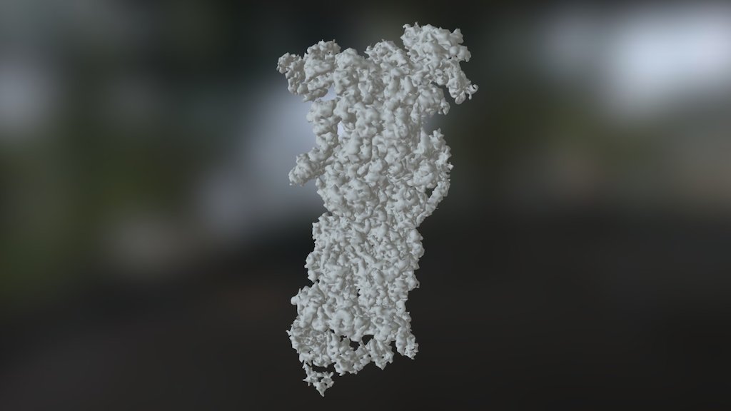
EMDB › EMD-3536 Single particle reconstruction achieved a remarkable 7.8Å resolution. 26S proteasome in presence of AMP-PNP (s3) was meticulously examined. Map released on 2017-03-08, marking a significant milestone. Overview of EMD-3536 reveals detailed insights into Saccharomyces cerevisiae's molecular machinery. Source organism: Saccharomyces cerevisiae (strain ATCC 204508 / S288c) [559292], a strain with a rich history of scientific exploration. Fitted atomic model: 5mpb, provides precise data for further analysis and research. Related EM entries by publication: EMD-3534, EMD-3535, EMD-3537 were published concurrently, shedding light on the proteasome's complex mechanisms. 3D bio-notes are available for this entry, a valuable resource for scientists worldwide. Primary publication: Structural insights into the functional cycle of the ATPase module of the 26S proteasome have been revealed through rigorous research and experimentation. Wehmer M, Rudack T, Beck F, Aufderheide A, Pfeifer G, Plitzko JM, Forster F, Schulten K, Baumeister W, Sakata E made significant contributions to the field of molecular biology. Their groundbreaking findings were published in Proc. Natl. Acad. Sci. U.S.A. 114 1305-1310 (2017), a testament to their dedication and expertise. PMID: 28115689 serves as a reference point for future research and discovery.
With this file you will be able to print EMDB › EMD-3536 with your 3D printer. Click on the button and save the file on your computer to work, edit or customize your design. You can also find more 3D designs for printers on EMDB › EMD-3536.