Dental Intra Oral X-ray Pos - question
sketchfab
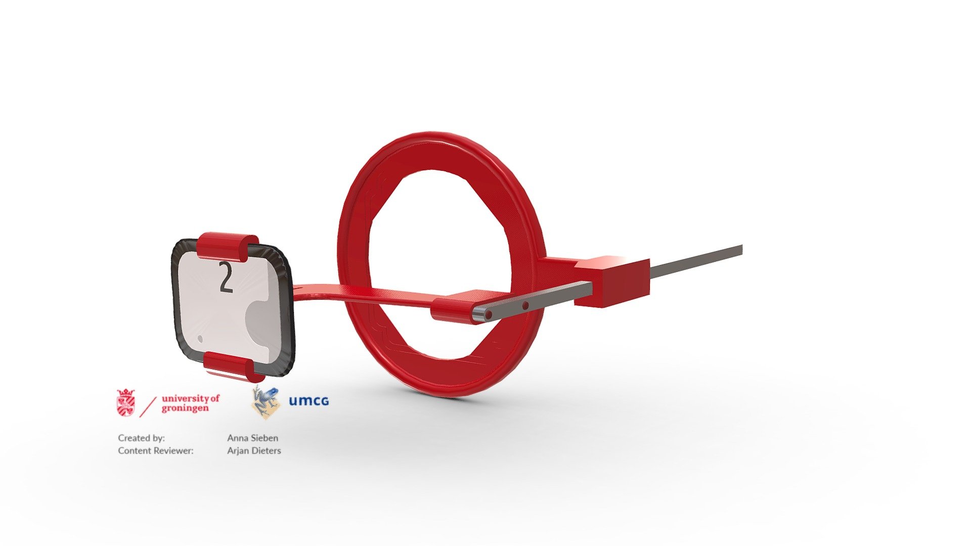
Dental X-rays are diagnostic tools that dentists use to assess oral health by capturing detailed images of teeth and gums with minimal radiation exposure. By evaluating these internal structures, dentists can identify issues like cavities, tooth decay, and impacted teeth. This illustration demonstrates the ideal positioning for a bite-wing X-ray, which showcases the upper and lower teeth in a specific region of the mouth. Each bite-wing image reveals a single tooth from its crown to the level of the surrounding bone. Bite-wing X-rays effectively detect decay between teeth and changes in bone density caused by gum disease. These X-rays also enable dentists to determine if a crown or other restorations fit properly, as well as monitor wear and breakdown of dental fillings. This model is featured in an online radiology manual designed for dental students.
With this file you will be able to print Dental Intra Oral X-ray Pos - question with your 3D printer. Click on the button and save the file on your computer to work, edit or customize your design. You can also find more 3D designs for printers on Dental Intra Oral X-ray Pos - question.